How I Tell Bacterial Leaf Spot vs Fungal Spots: Treat Tropical Plants In 5 Easy Steps
Learn how to distinguish water-soaked bacterial lesions from concentric fungal rings, why daily leaf misting spreads pathogens, and how to clinically treat infected tropical foliage.
The Misting Bottle Disaster
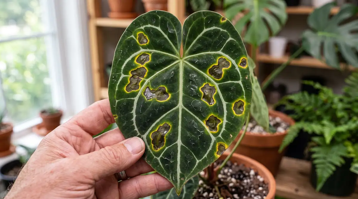
Early in my growing hobby, I acquired a gorgeous, velvety Anthurium clarinervium. Wanting to give it the tropical humidity it craved, I diligently misted its leaves twice a day with a spray bottle. Within three weeks, small, dark brown spots appeared near the leaf margins, each surrounded by a vivid yellow halo.
Assuming it was an ordinary fungal blight, I sprayed standard organic neem oil across the foliage. Over the next five days, the spots expanded rapidly, coalescing into translucent, greasy brown patches that smelled faintly foul. Entire leaves collapsed into mush.
That costly mistake led me to study plant pathology. I had misdiagnosed a bacterial infection caused by Xanthomonas as a simple fungal issue. By misting the plant daily, I had created a swimming pool for motile bacterial cells to enter open stomata and hydathodes. Understanding the exact microscopic distinctions between bacterial and fungal pathogens is essential to applying the correct chemical controls before valuable leaves are destroyed.
The Science: Pathogen Biology, Invasion Routes, and Lesion Morphology
1. Bacterial Leaf Spot Pathogens (Xanthomonas, Pseudomonas, Pectobacterium)
Bacterial pathogens are single-celled prokaryotic organisms that lack the mechanical structures needed to pierce intact leaf cuticles (Agrios, 2005). Instead, they enter plants through natural microscopic openings (stomata, hydathodes along leaf edges) or wounds caused by insect feeding and handling (Chase, 1987).
Once inside intercellular spaces, bacteria secrete pectolytic enzymes that dissolve cell walls, releasing cellular fluids. This breakdown creates the classic water-soaked, translucent appearance of bacterial lesions. Because bacterial spread within the leaf is physically blocked by major vascular bundles, bacterial spots often develop distinct angular borders constrained by veins.
2. Fungal Leaf Spot Pathogens (Colletotrichum, Cercospora, Alternaria)
Fungi are eukaryotic organisms that produce microscopic spores. When a fungal spore lands on a leaf surface in the presence of free water, it germinates and produces an infectious structure called an appressorium (Daughtrey et al., 1995). The appressorium exerts immense mechanical pressure to punch directly through the waxy plant cuticle.
As fungal hyphae radiate outward from the initial infection point, they produce distinctive concentric rings (often called target rings or bullseye patterns). The center of a fungal spot dries out into a crisp, papery texture and eventually develops tiny, raised black dots called pycnidia or fruiting bodies that house thousands of new spores.
3. The 3 Diagnostic Field Tests
You can identify the infection type in your own home using three simple observational tests:
- The Backlight Test: Hold the affected leaf up to a bright light source. Bacterial spots display a translucent, oily, water-soaked perimeter. Fungal spots appear opaque with dry, defined margins.
- The Lesion Geometry Test: Check whether the lesion stops abruptly at leaf veins. Angular spots that do not cross veins indicate bacterial infection. Circular spots with concentric growth rings indicate fungal infection.
- The Surface Texture & Magnifier Check: Touch the center of the spot. Dry, papery spots bearing pinpoint black dots under a 10x hand lens are fungal fruiting bodies. Mushy, greasy spots that occasionally exude a cloudy fluid droplet are bacterial colonies.
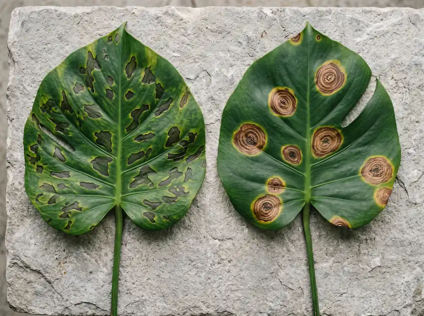
Bacterial Spot vs. Fungal Spot: Clinical Comparison
Use this diagnostic reference to quickly identify and treat foliar diseases:
| Diagnostic Trait | Bacterial Leaf Spot | Fungal Leaf Spot |
|---|---|---|
| Primary Pathogens | Xanthomonas, Pseudomonas, Pectobacterium | Colletotrichum (Anthracnose), Cercospora, Alternaria |
| Lesion Shape & Margins | Angular, sharply delimited by major veins | Circular or oval, concentric target rings |
| Tissue Appearance | Water-soaked, greasy, dark brown to black | Dry, papery, tan center with dark outer rim |
| Fruiting Bodies Present | None (Liquid bacterial ooze under humidity) | Yes (Pinpoint black pycnidia in lesion center) |
| Effective Chemical Treatment | Liquid Copper Octanoate (Copper soap bactericide) | Copper Fungicide, Sulfur, or Biofungicides |
5-Step Clinical Treatment Protocol
Step 1: Isolate the Infected Plant
Immediately move the symptomatic plant away from the rest of your collection to prevent splash transmission. Follow the isolation standards outlined in our guide on Quarantine Protocols for Indoor Tropicals.
Step 2: Surgical Excision of Severely Damaged Leaves
Sterilize a sharp pair of pruning shears with 70% isopropyl alcohol. Cut away leaves that have lost more than 40% of their surface area. For valuable specimens with only isolated spots, cut out the individual lesion along with a 1/4-inch border of healthy green tissue. Wipe your blade with alcohol between every single cut.
Step 3: Apply Liquid Copper Octanoate
Copper ions disrupt protein synthesis in both bacterial and fungal cells (Pscheidt & Ocamb, 2020). Spray a registered liquid copper soap bactericide/fungicide thoroughly over the upper and lower leaf surfaces. Repeat application every 7 to 10 days for three cycles.
Step 4: Cease All Overhead Spraying
Keep foliage completely dry. Water exclusively at the soil level or via bottom-watering trays. Pathogenic bacteria and fungal spores require a continuous film of free water for at least 4 to 6 hours to germinate and establish infection.
Step 5: Increase Canopy Airflow
Position an oscillating fan on low speed near your plants. Continuous gentle air movement rapidly evaporates any accidental droplets and prevents microclimates of saturated air from lingering around leaf stomata.
Troubleshooting Leaf Spot Inquiries
• Will Damaged Leaves Turn Green Again After Treatment?
No. Necrotic leaf spots represent permanently dead plant cells. Successful treatment stops the spots from expanding and protects emerging new leaves from becoming colonized.
• Can Neem Oil Treat Bacterial Leaf Spot?
Neem oil functions primarily as an insecticide and mild anti-sporulant for specific surface fungi, but it lacks bactericidal efficacy against systemic bacterial pathogens like Xanthomonas. Use copper-based bactericides or biological Bacillus inoculants for bacterial problems.
• Why Do Leaf Spots Always Have a Yellow Halo?
The yellow halo (chlorosis) is caused by phytotoxins secreted by the invading pathogen as it advances, breaking down chlorophyll ahead of the necrotic cellular collapse.